The neural tube is the precursor of the central nervous system which is the brain and spinal cord. Neurulation is the process in which the formation of the neural tube takes place between day 16th until day 30th of gestation in humans.
Onset Of Development
The development of an embryo begins with an ovum and a sperm merging their nuclei together. Subsequently these cells divide and multiply into a packed group of cells called morula and the division continues as the morula furthers its cell division into a blastocyst. At this stage, the blastocyst comprises of 3 germinal layers starting from the inner most layer endoderm, mesoderm and the outer layer, ectoderm.
Onset Of Development Leading To Trilaminar Layer
The specification of germinal cells is also known as gastrulation. A flat epithelial sheet comprising of an outer layer which is the ectodermal tissue and an inner layer which is the neuroepithelium undergoes rostral and caudal elongation. Furthermore, this elongation process is coupled with conversion of cells towards the midline, a process known as convergent extension.
Neurulation
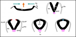
Neurulation proceeds with the bending of the neuroepithelium at the midline MHP (medial hinge point) to form apposing neural folds. The lateral edges of the neural folds bend DHLP (dorsolateral hinge point), and subsequently, the edges make contact, adhere and fuse at the dorsal midline. Neurulation completes at the 17 somites stage in the cranial region and the extreme end of spinal neural tube, posterior neuropore, closes at the 30 somites-stage.
Neurulation And Failure Of Neural Tube Closure
Any failure of closure at any stage of neurulation results in neural tube defects (NTDs). Exencephaly is a condition when the cranial neural tube fails to close leaving the neural folds splayed open, the brain later undergoes secondary degeneration that results in the exposed skull base. A more developed form of exencephaly, anencephaly exhibits a bare skull base lined by vascular tissue with little brain tissue left despite of normal neuronal growth during early gestation. Another form of NTD is spina bifida that occurs when the spinal neural tube fails to close and has lesser death prevalence but individuals with spina bifida may suffer from incontinence and defect in motor and sensory innervation in the legs. Hydrocephalus and vertebral curvature or more commonly known as scoliosis are associated with NTD but is not per se a representation of failure of neural tube closure. The severity of the condition is dependent on the location of the lesion. It appears that the higher the lesion on the spinal column, the more severe the condition. The most severe form of NTD is craniorachischisis whereby the neural tube of the cranial and entire spinal region fails to close.
NTDs are the second leading cause of congenital birth defects in humans with the incidence of 1/1000 in American Caucasian and 0.5-2 per 1000 pregnancies worldwide. Studies were done on maternal supplementation to prevent NTDs. Among the supplements that were given periconceptionally were folic acid, methionine and inositol.
Mechanism Of Neural Tube Closure.
Se: Surface ectoderm
Ne: Neuroepithelium
Me: Mesoderm
MHP: Median hinge point
DHLP: dorsolateral hinge point
POF: point of fusion
Nt: notochord.
- A) A flat sheet of neural plate elongates anterior posteriorly and narrows toward the midline
- B) The neural plate begins to elevate forming a MHP at the midline
- C) The opposing fold bends to form DHLP
- D) The opposing fold, closing each other to adhere and fuse
- E) The neural tube undergoes remodelling and the surface ectoderm separates from the neural tube.
